

(Editor’s Note: This is a Guest Post on an ultrasound case submitted to me by a 3rd-year medical student (Danielle Hamilton) who was able to use POCUS on her internal medicine rotation. I think this is a great case illustrating how even a medical student with a simple handheld ultrasound device can use POCUS on their rotations. This further highlights the importance of learning POCUS as trainees and students are becoming very adept at this skill!
If you have any interesting ultrasound cases or topics that you would like to share with thousands of other POCUS enthusiasts read our Guest Post Policy and feel free to contact me at Editor@Pocus101.com)
Authors: Danielle Hamilton and Derek Chinn
Intro
Bedside ultrasound is a useful, non-invasive imaging modality that can be used to easily assess left ventricular function. In this post, we discuss the case of an elderly male patient with acute-on-chronic systolic heart failure as well as the clinical significance, indications, and techniques of bedside cardiac ultrasound in the measurement of left ventricular ejection fraction (LVEF).
Case
An 87-year-old man with a past medical history significant for hypertension, hyperlipidemia, and chronic systolic heart failure secondary to non-ischemic cardiomyopathy presented to the emergency department for acute-onset altered mental status. Vitals revealed tachycardia and hypotension to the 80s systolic. Fluid status was assessed with bedside ultrasound and revealed clear lungs, an LVEF of 35-40%, and a large inferior vena cava (IVC) that did not collapse with inspiration. Due to suspicion of distributive shock, a trial 500-mL bolus of Lactated Ringers (LR) was administered with temporary improvement in blood pressure. Telemetry showed an abnormal rhythm, so a STAT EKG was ordered and revealed new-onset atrial fibrillation (A-fib) with RVR. IV metoprolol was administered but did not resolve the A-fib with RVR. Vasopressors and an amiodarone drip were initiated in the emergency department.
After stabilization, the patient was admitted to the medical intensive care unit (MICU) and managed with antibiotics, fluids, antiarrhythmics, and vasopressors. The patient remained afebrile, and infectious workup was unremarkable. He then developed lower extremity edema, jugular venous distention, and cold extremities on physical exam but denied chest pain and shortness of breath. His vitals showed hypotension to the 60s systolic and tachycardia to the 120s. A STAT EKG was unremarkable for ST changes, although his troponins were elevated above baseline, consistent with non-ST segment elevation myocardial infarction (NSTEMI). Due to the presence of cardiogenic shock, he was started on dobutamine and furosemide drips. His volume status significantly improved, hemodynamics stabilized, and he was weaned to room air.
The patient was then downgraded to the internal medicine hospitalist service for further management of acute-on-chronic systolic heart failure. A repeat bedside ultrasound was performed by a third-year medical student using a handheld Butterfly probe and an iPhone (see figures below). At this time, LVEF was estimated to be severely depressed (<30%) in the parasternal long-axis (PLAX) view. The patient was discharged to SNF in stable condition with Tylenol, amiodarone, apixaban, carvedilol, and furosemide and instructions to follow up with his primary care physician for further management of his systolic heart failure.
Figure 1: PLAX view of systolic ejection. Note the poor squeezing motion of the left ventricle during systole.

Figure 2: PLAX view of diastolic filling. The tip of the anterior mitral valve leaflet (AML) is indicated by the green arrow. Note the limited motion of the AML during diastole.

Discussion
Bedside cardiac ultrasound is useful for assessing systolic function via estimation of LVEF (1, 2, 3, 4, 5). Indications for performing bedside cardiac ultrasound include chest pain, shortness of breath, hypotension, chest trauma, and cardiac arrest (2). The patient reported in this case had acute-on-chronic systolic heart failure with reduced LVEF and subsequently went into cardiogenic shock. A third-year medical student obtained the PLAX view and estimated this patient’s LVEF at the bedside using a Butterfly ultrasound probe and an iPhone. This estimation was made based upon the poor squeezing motion of the left ventricle (LV) during systole and the limited motion of the anterior mitral valve leaflet toward the interventricular septum (IVS) during diastole.
(Editor’s note: for a FULL tutorial on how to evaluate left ventricular ejection fraction click HERE)
Table 1: LVEF classifications based upon LV squeezing during systole and AML motion during diastole (1).
| Classification | LVEF |
| Hyperdynamic | ≥70% |
| Normal | 55-69% |
| Mildly reduced | 45-54% |
| Moderately reduced | 30-44% |
| Severely reduced | <30% |
The eyeball method has been shown to underestimate the LVEF by 8.4% on average, even amongst experienced clinicians (3). It is thus recommended that estimations of LVEF using the eyeball method can be confirmed using quantitative measurements of LVEF (3). Additionally, it is recommended to obtain multiple views in order to obtain a more accurate estimation of LVEF (3). Quantitative measurements such as E-point septal separation (EPSS), fractional shortening, fractional area exchange, and Simpson method more accurately estimate the LVEF when obtained by an experienced user (1). For example, an EPSS of >7 mm measured in the PLAX view has been shown to be 100% sensitive for LVEF <30% (4). It is important to note that wildly false values are possible with quantitative measurements if a slightly incorrect angle or cut is obtained (1). This limitation increases the utility of the eyeball method which can be easily performed and fairly accurately interpreted by a novice user (1, 3).
The four key cardiac views include parasternal long axis (PLAX), parasternal short axis (PSAX), apical 4-chamber (A4C), and subxiphoid (2). In this case report, we highlight the PLAX view. To obtain this view, the ultrasonographer should ask the patient to lay supine or in the left lateral decubitus position. The probe should be placed at the 3rd, 4th, or 5th left intercostal space (LICS) immediately adjacent to the sternum (5). The indicator on the probe should point toward the patient’s right shoulder, which is where the base of the heart lies (5). Cardiac mode should be selected, which can be confirmed by looking at the position of the indicator on the screen. Cardiac mode is unique from other modes in that the indicator on the screen is located on the right instead of the left (Figure 3).
(Editor’s note: for a FULL tutorial on how to perform Cardiac Ultrasound click HERE)
Figure 3: Cardiac mode. The blue arrow is pointing toward the indicator on the screen, which is located on the right.
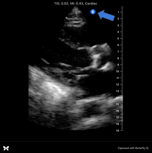
Figure 4: Labeled structures in the PLAX view. AV = aortic valve, DA = descending aorta, IVS = interventricular septum, LA = left atrium, LV = left ventricle, MV = mitral valve, LVOT = left ventricular outflow tract, P = pericardium, RV = right ventricle.

Conclusion
This case report describes an 87-year-old man with acute-on-chronic systolic heart failure. The clinical significance and indications of bedside cardiac ultrasound in qualitatively estimating LVEF as well as the method of obtaining the PLAX view are discussed.
(Editor’s Comments: This is a simple case showing how you can estimate LV Ejection Fraction and integrate it with your clinical reasoning. A couple of points here to emphasize. 1) When doing cardiac POCUS it may also be beneficial to add the lung ultrasound views to see if there is any pulmonary interstitial edema. This is especially useful in heart failure patients and when you are considering giving them IV fluid. 2) Once you are familiar with estimating ejection fraction it can also be helpful to measure the diastolic function, cardiac output, as well as venous congestion of your patients. This will give you an even more comprehensive understanding of the cardiac pressures and fluid status of the patient allowing you to more precisely start vasopressors, fluid administration, or diuresis.)
Works Cited
- Dinh, Vi, et al. Assessing Left Ventricular Ejection Fraction With Echocardiography. www.pocus101.com/assessing-left-ventricular-ejection-fraction-with-echocardiography/.
- Herbst MK, Velasquez J, O’Rourke MC. Cardiac Ultrasound. [Updated 2020 Jun 2]. In: StatPearls [Internet]. Treasure Island (FL): StatPearls Publishing; 2020 Jan-. Available from: https://www.ncbi.nlm.nih.gov/books/NBK470584/
- Holloway, C.J., Rider, O.J., Edwards, L.M. et al. Eyeball assessment of left ventricular function, compared to a quantitative assessment by cardiac magnetic resonance. J Cardiovasc Magn Reson 12, P251 (2010). https://doi.org/10.1186/1532-429X-12-S1-P251
- McKaigney CJ, Krantz MJ, La Rocque CL, et al. E-point septal separation: a bedside tool for emergency physician assessment of left ventricular ejection fraction. Am J Emerg Med. 2014; 32(6):493-497.
- Timothy Jang, M. (2020, December 05). Cardiac Evaluation using Bedside Ultrasonography: Practice Essentials, Positioning, Technique. Retrieved January 16, 2021, from https://emedicine.medscape.com/article/104401-overview


Great job!!! This is an excellent study.
Thanks Kim!!! Just spreading the echo knowledge to others just like you have taught me!! Appreciate the kind words.
Awwww, thank you so much!! What an honor. 😊😊
It was a great post thanks for contributing!
Thank you very much, because you give us great knowledge!
Thanks Thi! Appreciate the comment.