
Primary Authors: Jessica Ahn, Vi Dinh; Co-authors: Jade Deschamps, Satchel Genobaga, Annalise Lang, Victor Lee, Reed Krause, Devin Tooma, Seth White. Oversight, Review, and Final Edits by Vi Dinh (POCUS 101 Editor).
Revisions by: Jessica Kim and Caleb Tan
Ocular complaints are common in medicine and getting the correct diagnosis is crucial in saving your patient’s vision. However, fundoscopic eye exams using an ophthalmoscope are frustratingly difficult to perform and can miss ocular emergencies.
Point of care Ocular/Eye Ultrasound is a quick and non-invasive way to evaluate for the most common ophthalmic pathologies with very high sensitivity and specificity (Blaivas et al).
In this post, we will show you a step-by-step approach on how to use Ocular/Eye Ultrasound to:
- Perform an ultrasound exam of the eye
- Learn Ocular Ultrasound Anatomy
- Detect the most common atraumatic and traumatic eye pathologies
- Estimate intracranial pressure
After learning these principles, you will be able to use Point of Care Ocular Ultrasound to tackle any ocular complaints with ease! In addition, we made an Ocular Ultrasound PDF Pocket Card you can download as well.
Table of Contents
Ocular Ultrasound POCKET CARD PDF
Download Ocular Ultrasound Protocol Card PDF HERE

Pathology Illustrations adapted from ddxof: Ocular Ultrasound by Tom Fadial, CC BY-SA 4.0
Like this Post?
Sign Up For POCUS 101 Updates!

Indications and Contraindications
Ocular Ultrasound Indications
- Vision Loss
- Change of Vision
- Acute Eye pain
- Ocular Trauma
- Intraocular Foreign Body
- Suspected Elevation in Intracranial Pressure
Ocular Ultrasound Contraindication
The main contraindication to performing ophthalmic ultrasound is if a patient has a globe rupture. It is advised to refer any of these suspected patients immediately to an opthalmologist. However, some resources state ocular ultrasound may cautiously be performed if there is a copious amount of gel placed and no pressure is applied to the eye (Kilker 2014). Please use your institutional guidelines.
Ocular Ultrasound Preparation
Patient Preparation
- The patient may be fully supine or head of the bed can be elevated up to 45°
- Optional: Place a Tegaderm patch over the closed eye of interest
- Apply a generous amount of ultrasound gel on the closed eye or on top of the Tegaderm patch. This ensures the ultrasound probe does not contact or put too much pressure on the patient’s eye

Ocular Ultrasound Machine Preparation
- Transducer: Linear Ultrasound Probe
- Preset: Ocular (B scan) or Superficial Preset
- Depth: Approximately 4 cm
- Ocular Ultrasound Machine Placement: Place the ultrasound machine on the patient’s right side so you can scan with your right hand and manipulate ultrasound buttons with your left hand

Step-by-Step Ocular Ultrasound Protocol
In this section, you will learn a step-by-step approach on how to use ultrasound of the eye to detect normal ocular structures in the transverse and sagittal views, assess for extra-ocular movements, and measure the optic nerve sheath diameter to estimate intracranial pressure.
Step 1: Anchor the Probe
In addition to a generous amount of gel, it is important to anchor your probe to decrease the amount of pressure applied to the patient’s eyes.
- Grasp the linear probe and anchor your fingers on a bony surface of the patient’s face.
- The example below assumes using the right hand to scan the patient.
- For the Right eye, anchor your right pinky finger on the patient’s nose.
- For the Left eye, anchor your right pinky finger or palm on the zygomatic arch.


Step 2: Obtain Transverse View
- Place the probe lightly on the gel covering the patient’s eye with the probe indicator pointed towards the patient’s right to obtain a transverse view.

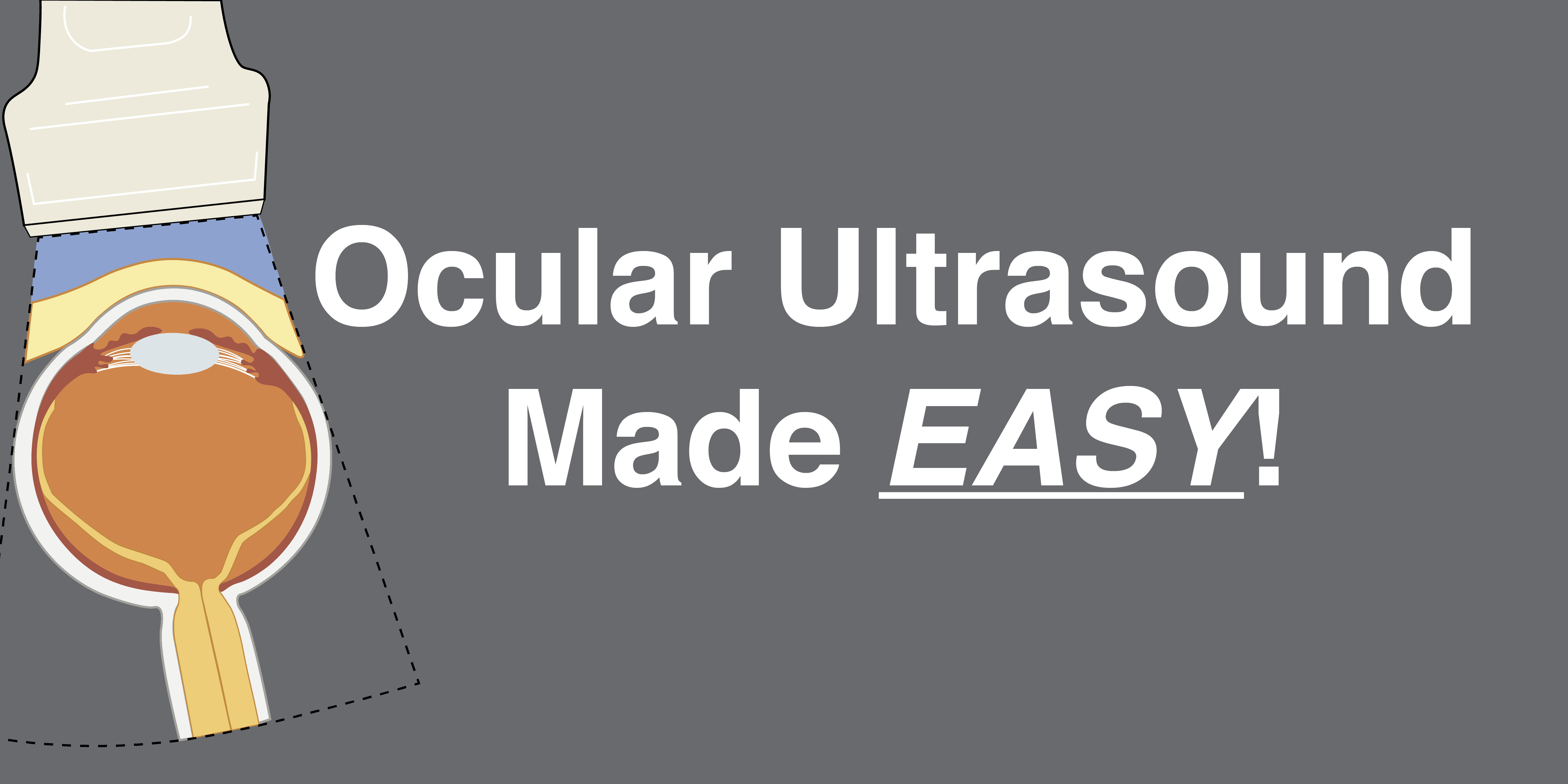
Identify the following ocular ultrasound anatomy from anterior to posterior:
- Eyelid
- Anterior Chamber
- Lens
- Iris
- Vitreous Body
- Retina
- Optic Nerve
- *Make sure to tilt/fan through the entire eye


Assess for Extraocular Movements
- Next, ask the patient to look left and right to evaluate for extraocular movements. This is important when patients have severe periorbital edema from facial trauma.
- In the transverse view, you are looking for MEDIAL and LATERAL (or Left and Right) movements of the eye.
- Increase the gain slowly to better detect intraocular pathologies such as mobile findings of retinal detachment, posterior vitreous detachment, or vitreous hemorrhage.

Step 3: Obtain Sagittal View
- Next, turn the probe 90° clockwise so the indicator points superiorly towards the patient’s head to obtain a sagittal view. Identify the same structures you found in the transverse view.

- Then, have the patient look up and down and increase the gain slowly to assess for symmetric extra-ocular movements and to rule out intraocular pathology.
- In the sagittal view, you are looking for Superior and Inferior (or Up and Down) movements of the eye.
Step 4: Measure Optic Nerve Sheath Diameter (ONSD)
The Optic Nerve Sheath Diameter (ONSD) is an important measurement that can be used to detect elevated intracranial pressure (ICP).
- In the transverse view, rock the probe about 10-15° laterally to visualize where the hypoechoic (darkly colored) optic nerve radiates away from the base of the globe (see figure below).
- Tip: If you do not see the ocular nerve immediately, tilt the probe up and down until it comes into view.
- Once you have a good view, freeze the image.


- Next, use the calipers again to measure the outermost lateral borders of the optic nerve sheath (anechoic border). The figure below measures the ONSD diameter to be 4.68mm (.468cm).
- To obtain better accuracy, you can obtain a few measurements and take the average of the ONSD values.

Interpretation of Optic Nerve Sheath Diameter (ONSD)
- In adults, an ONSD < 5 mm indicates that the patient has a normal optic nerve width and a normal intracranial pressure (ICP) value of <20 cm H2O. However, if the ONSD > 5 mm, you will not be able to correlate this with an exact ICP value; all this tells you is that the ICP is elevated (Kilker, et al).
- Below is a quick table regarding normal ONSD measurements for adults vs children:
Editor’s Note: To remember how to measure ONSD, think of a 3×5 index card. You need to measure 3 mm posterior to the globe and >5 mm indicates a high ICP.
| Age Category (Years) | Normal ONSD Measurement |
| Adults (16+) | < 5 mm |
| Children (1-15) | < 4.5 mm |
| Children (<1) | < 4 mm |

Video Summary of Ocular Ultrasound
Here is a brief video summarizing how to do the ocular ultrasound exam:
Like this Post?
Sign Up For POCUS 101 Updates!
Ocular Ultrasound Pathology Overview
In this section, we will go over the ocular pathology related to the 3 most common ocular presentations for vision change:
- Painless Vision Loss
- Traumatic Vision Loss
- Elevation of Intracranial Pressure (Intracranial hemorrhage and Pseudotumor Cerebri)
The Ocular Ultrasound PDF Pocket Card below can be used as a reference guide as you go through the different ocular pathologies (click HERE to download the PDF):
Painless Vision Loss
Retinal Detachment (RD)
A retinal detachment is defined by a separation of the sensory retina from the underlying retinal pigment epithelium. This cuts off the blood supply to the rods and cones in the eye and can cause permanent vision loss. A retinal detachment is an ocular emergency that must be referred immediately to an ophthalmologist (Ghazi & Green).
A patient with a detached retina typically presents with painless, fixed visual field loss, new floaters, or flashes (photopsia) with the perception of a curtain coming down (inferior detachment) or up (superior detachment). Unfortunately, fundoscopy using a direct ophthalmoscope is limited and can miss retinal detachments.
There are three major types of retinal detachment (rhegmatogenous, traction, and exudative):

Ocular ultrasound is both sensitive and specific for diagnosing retinal detachments. While distinguishing between the 3 types of retinal detachment is important from a treatment standpoint, ocular ultrasound should not focus on distinguishing between them. The primary purpose of POCUS is to be able to diagnose a retinal detachment and then allow the ophthalmologist to help definitively diagnose the retinal detachment type and treatment option.
Retinal Detachment Ultrasound Findings
On B scan (B mode), ocular ultrasound retinal detachment is best visualized in normal and often low gain settings. A retinal detachment looks like a thick/hyperechoic membrane lifted off of the posterior surface of the globe that floats and moves with the patient’s eye movement.
If the retinal detachment is large, the hyperechoic membrane will be tethered to the optic nerve. However, if the retinal detachment is small or not near the optic nerve, the hyperechoic membrane will be tethered closely to the back wall of the eye and will not move much with eye movement. It is important to scan the eye in multiple axes (transverse and sagittal) to detect small retinal detachments.

See below for an ocular ultrasound retinal detachment image and video. Notice how the retinal detachment is tethered to the optic nerve as the patient moves their eye.


Posterior Vitreous Detachment (PVD)
A posterior vitreous detachment (PVD) occurs when the vitreous body separates from the posterior portion of the normal retina, but the retina is still intact.
There is a higher prevalence of PVD in elderly and myopic patients. Patients with posterior vitreous detachment (PVD) often present with fluctuating cloudy vision, acute floaters, and brief flashes (photopsia).
Posterior Vitreous Detachment Ultrasound Findings
On ocular ultrasound, posterior vitreous detachment looks like a thin, hyperechoic membrane lifted off the posterior surface of the globe that is NOT tethered to the optic nerve. The membrane will freely move with ocular movements in an undulating fashion, like “swaying seaweed.”


Differentiating RD and PVD with Ocular Ultrasound
Retinal detachment and posterior vitreous detachment can present with similar symptoms but the management and prognosis between the two conditions are very different.
While retinal detachment is an ocular emergency, posterior vitreous separation is generally not. Ultrasound of the eye can help diagnose and distinguish between Retinal Detachment and Posterior Vitreous Detachment but an urgent/emergent ophthalmology consult should be placed to definitively differentiate between the two.
If you see a hyperechoic membrane tethered to the optic nerve, this is a retinal detachment. However, if the hyperechoic flap is along the posterior wall of the eye and not tethered to the optic nerve, this could be a retinal detachment or a posterior vitreous detachment.
The table below shows the differences between retinal detachment and posterior vitreous detachment:
| Retinal Detachment | Posterior Vitreous Detachment | |
| Emergency Referral Needed | Yes | No |
| Vision Changes | Constant vision loss | Fluctuating vision blur |
| Optic Nerve Sheath Attachment | Hyperechoic line most often attached to the optic nerve sheath (unless RD is not right next to optic nerve) | Hyperechoic line is not attached to the optic nerve sheath |
| Mobility with eye movements | Less mobile: adheres closely to wall | Very mobile; undulates like “seaweed” |
| Thickness | Thick folded membrane | Thinner, smooth folded membrane |
Vitreous Hemorrhage (VH)
Vitreous hemorrhage occurs when extravasated blood is inside or around the vitreous humor of the eye. This can occur from damage to normal blood vessels, growth of abnormal blood vessels, or bleeding from other parts of the eye.
Symptoms of vitreous hemorrhage include sudden, painless vision loss, photophobia, and the perception of shadows and cobwebs floating in front of their eyes. Sometimes, patients describe their vision as worse in the morning because blood has settled at the back of their eye during the night.

Vitreous Hemorrhage Ultrasound Findings
On ophthalmic ultrasound, vitreous hemorrhage looks like echogenic material in the posterior chamber. If you ask the patient to move their eye on ophthalmic ultrasound, vitreous hemorrhage is best visualized with normal or high gain settings. Vitreous hemorrhage looks like echogenic material in the posterior chamber. If you ask the patient to move their eye side-to-side, you may see the washing machine sign, where the echogenic material appears to swirl like clothes in a washing machine.


Central Retinal Artery Occlusion (CRAO)
The central retinal artery (CRA) is a branch of the ophthalmic artery that travels within the optic nerve to supply blood to the retina. Occlusion of the central retinal artery is an ophthalmic emergency as the remaining posterior ciliary artery cannot sufficiently supply the retina’s needs. Emergent Opthalmology consult must be placed and hyperbaric oxygen therapy may be required.


CRAO commonly occurs from carotid artery atherosclerosis and plaque embolism. Some contributing risk factors for include diabetes, hypertension, cardiovascular disease, and hyperlipidemia (Varma et al).
Patients with CRAO usually have sudden, painless monocular vision loss. If you suspect CRAO, it is important to measure the blood pressure as there is a strong relationship between CRAO and hypertension (Varma et al). Another important area to assess is the radial pulse rate and rhythm. Atrial fibrillation has a high risk for embolisms and can be detected as an irregularly irregular pulse.
Central Retinal Artery Occlusion (CRAO) Ultrasound Findings
CRAO is a rare finding and requires color Doppler mode to diagnose with ocular ultrasound. On the ocular ultrasound exam, you will find diminished or absent flow of the central retinal artery (see below). You may also only see flow from the central retinal vein (blue on color Doppler) with absent central retinal arterial flow (red on color Doppler)
(Editor’s Note: In some circumstances, you may also still see some arterial flow on color Doppler from the posterior ciliary artery, even though the central retinal artery is occluded)


Intraocular Masses
Intraocular masses are rarely detected and diagnosed in the Emergency Department (ED). Masses can be benign or malignant with retinoblastoma being the most common primary malignant intraocular tumor in kids and melanoma in adults (Bornfield, Loe). When fundoscopic exams are limited, ultrasound provides an easy, accessible way to diagnose ocular masses. Patients with intraocular masses can present with symptoms of headache, neurological complaints, and vision loss and changes.
Ocular ultrasound has significant value when time and ocular equipment are limited especially in the ED (Blavias). Additionally, findings such as severe periorbital swelling may limit or prevent a fundoscopic exam.
In one case report on ocular melanoma, a patient presented with headaches along with hearing and visual deficits. Her fundoscopic exam demonstrated decreased visual acuity and red reflex, but the rest of the exam was limited. US was used and showed a hypoechoic mass in the right posterior eye, which led to an orbital CT and ophthalmology consult and admission (Loe).
Intraocular Mass ultrasound findings:
On ocular ultrasound, you will visualize a mass that can present with various echogenicities. Retinoblastoma often show microcalcifications (Brennan et al 2012). Whenever performing ocular ultrasound, only minimal pressure should be applied to avoid increases in intraocular pressure and vagal-type responses.

Like this Post?
Sign Up For POCUS 101 Updates!
Traumatic Vision Loss
Retinal Detachment
Traumatic Retinal Detachments will have similar ocular ultrasound findings to atraumatic retinal detachments as described above.

Intraocular Foreign Body (IOFB)
Foreign bodies can enter the eye and lead to globe rupture, infection, retinal toxicity, and vision loss if not removed (Pinto et al).
These patients often present with a painful foreign body sensation in the eye, decreased vision, redness, tearing, flashes, or floaters.
Intraocular Foreign Body Ultrasound Findings
On ocular ultrasound, you can find a bright, hyperechoic object with an associated reverberation artifact (if object is metallic).


Globe Rupture
Globe rupture occurs when a blunt or penetrating trauma disrupts the integrity of the outer membranes of the eye. Traumatic globe rupture is a major ophthalmologic emergency and must be referred to ophthalmology immediately.
These patients present with painful vision loss and extrusion of vitreous fluid.

(Editor’s Note: Some may say globe rupture is a relative contraindication to ocular ultrasound. Use extreme care and copious amounts of gel if you are attempting ultrasound on a patient with suspected globe rupture. Any additional pressure on the eye from the transducer can worsen extrusion of the vitreous humor.)
Globe Rupture Ultrasound Findings
On ocular ultrasound, you may find buckling of the sclera, asymmetric loss of the normal spherical shape of the globe, decreased size of globe, flattening or compression of the anterior chamber, or often have vitreous hemorrhage (Kilker et al).


Lens Dislocation
Lens dislocation typically occurs after blunt or penetrating trauma and can be partial (subluxation) or complete lens dislocations.
These patients often present with a change in vision (blurring, double vision, seeing the edge of the lens) and normal pupillary response.
Lens Dislocation Ultrasound Findings
On ocular ultrasound, you will see the lens as a bi-convex structure with hyperechoic borders floating posteriorly in the vitreous body.


Retrobulbar Hemorrhage
Retrobulbar hemorrhage is a rare finding that occurs when blood accumulates in the retrobulbar space, often from orbital trauma or postoperative complications from eye surgery.
This condition is an ocular emergency as the quick elevation of retro-orbital pressure can cause reduced circulation, ischemia, and finally necrosis of the optic nerve with irreversible blindness (Roque et al).
Patients often present with sudden-onset pain, proptosis, conjunctival edema, or limited extra-ocular movements.
Retrobulbar Hemorrhage Ultrasound Findings
On ocular ultrasound, you may see a “Guitar Pick Sign” where the increased pressure from the retrobulbar hematoma distorts the spherical globe into a conical shape (Kilker et al).


Periorbital Edema
Certain types of trauma can cause significant enough periorbital edema that it can be challenging to retract the eyelids of patients. It is important to assess extraocular eye movements to detect muscular entrapment or cranial nerve defects (Harries et al).


Like this Post?
Sign Up For POCUS 101 Updates!
Evaluation of Elevated Intracranial Pressure/Papilledema
Elevated intracranial pressure (ICP) may be a sign of a serious or life-threatening medical condition and it may be helpful to augment the fundoscopic exam with ultrasound to evaluate for papilledema which can be from a chronic increase in intracranial pressure.
Intracranial Pressure Evaluation with Ocular Ultrasound
Ultrasound of the optic nerve sheath diameter has proven to be a reliable, non-invasive, and rapid method to estimate ICP measurement. Any elevation in ICP causes distension of the ocular nerve sheath (>5mm) likely within minutes, making it a prime way to measure ICP in emergent situations (Rajajee et al).
Papilledema Evaluation with Ocular Ultrasound
If the ICP is chronically elevated, the cerebrospinal fluid will accumulate within the enlarged optic nerve and cause the optic disc to be raised and bulge into the retina. The combination of a widened ONSD along with optic disc edema is known as papilledema.
On ocular ultrasound, papilledema presents with an ONSD > 5 mm and an optic nerve disc bulging of more than 0.6 mm (Teismann et al). This differs from an acute elevation in ICP which only shows an ONSD > 5 mm on ultrasound.
Papilledema can be caused by brain masses, hydrocephalus, stroke or idiopathic intracranial hypertension/pseudotumor cerebri. The presence of unilateral papilledema can signal rare etiologies such as optic neuritis.
Differences between Elevated ICP, Papilledema, and Pseudopapilledema
It is important to note that optic disc edema doesn’t always correlate with chronic ICP elevation leading to papilledema. Pseudopapilledema is a benign elevation of the optic nerve head that has no related elevation in ICP. This condition often occurs if there is a small crowded optic nerve head, tilted optic disc, or optic nerve head drusen (Chiang et al).
Below is a table showing the differences between elevated ICP, Papilledema and Pseudopapilledema.
| Acutely Elevated ICP | Papilledema | Pseudopapilledema | |
| ICP | Acutely high | Chronically high | Normal |
| ONSD | > 5 mm | > 5 mm | Normal |
| Optic Disc Bulging | Normal | > 0.6 mm | > 0.6 mm |
| Example | Early intracranial hemorrhage | Pseudotumor cerebri | Small crowded optic nerve head |
In the upcoming sections, we will focus on the two most common pathologies of increased ICP and/or papilledema: intracranial hemorrhage and pseudotumor cerebri (idiopathic intracranial hypertension).
Intracranial Hemorrhage
Intracranial hemorrhage occurs when a blood vessel within the skull ruptures or leaks. This condition can occur with physical trauma from a head injury or a ruptured aneurysm. The rapid build-up of blood can increase the ICP so quickly that permanent brain damage can occur if the pressure is not alleviated.
Oftentimes, these patients are comatose and will need emergent lowering of their ICP using pharmacologic (mannitol and hypertonic saline) or neurosurgical decompression.
Intracranial Hemorrhage Ultrasound Findings
On ocular ultrasound, you will find elevated ONSD measurements > 5 mm. Because ultrasound scanning for this condition is often done soon after trauma, there may not be enough time for the elevated ICP to progress to papilledema. Therefore, it is unlikely to see optic disc bulging from an acute intracranial hemorrhage on ultrasound. This contrasts with chronically increased intracranial pressure where the optic nerve swelling can be visualized with ultrasound.


Pseudotumor Cerebri (Idiopathic Intracranial Hypertension)
Pseudotumor cerebri syndrome occurs when there is raised intracranial pressure but no known etiology (Mollan et al).
These patients present with headaches that are typically worst in the morning when they wake up or when they are lying down (Chiang et al). Some other symptoms can include transient vision loss, diplopia, pulsatile tinnitus, nausea, or vomiting. Idiopathic intracranial hypertension (IIH) predominately affects overweight women of childbearing age (Chiang et al).
This disorder needs to be rigorously monitored to ensure the ICP is not elevating to levels dangerous enough to cause visual loss. Neurology consult and lumbar puncture are recommended after brain imaging to rule out any brain abnormalities. Treatment with a lumbar puncture to remove excess CSF has been shown to significantly reduce the mean ONSD, indicating that this procedure can lower the ICP to normal values (Bauerle & Nedelamnn).
Pseudotumor Cerebri (Idiopathic Intracranial Hypertension) Ultrasound Findings
On ocular ultrasound, you will find elevated ONSD measurements > 5 mm. Because this is typically a chronic condition, patients often have signs of Papilledema with optic disc bulging/elevation.


References
- Blaivas M, Theodoro D, Sierzenski PR. A study of bedside ocular ultrasonography in the emergency department. Acad Emerg Med 9, 791–799 (2002).
- Baker, N. et al. Can emergency physicians accurately distinguish retinal detachment from posterior vitreous detachment with point-of-care ocular ultrasound? Am J Emerg Medicine 36, 774–776 (2018).
- Bäuerle J, Nedelmann M. Sonographic assessment of the optic nerve sheath in idiopathic intracranial hypertension. J Neurol 258, 2014–9 (2011).
- Case courtesy of Dr Maulik S Patel, Radiopaedia.org. From the case rID: 10009
- Chiang J, Wong E, Whatham A, et al. The usefulness of multimodal imaging for differentiating pseudopapilloedema and true swelling of the optic nerve head: a review and case series: Differentiating optic nerve head swelling. Clin Exp Optom 98, 12–24 (2014).
- The Eye Institute. https://www.eyeinstitute.co.nz/about-eyes/a-to-z-of-eyes/conditions/vitreous-haemorrhage
- Ghazi, N. G. & Green, W. R. Pathology and pathogenesis of retinal detachment. Eye 16, 411–421 (2002).
- Hansen, H. & Helmke, K. The subarachnoid space surrounding the optic nerves. An ultrasound study of the optic nerve sheath. Surg Radiol Anat 18, 323–328 (1996).
- Harries, A., Shah, S., Teismann, N., Price, D. & Nagdev, A. Ultrasound assessment of extraocular movements and pupillary light reflex in ocular trauma. Am J Emerg Medicine 28, 956–959 (2010).
- Mollan SP, Ali F, Hassan-Smith G, et al. Evolving evidence in adult idiopathic intracranial hypertension: pathophysiology and management. J Neurology Neurosurg Psychiatry 87, 982–92 (2016).
- Kilker, B. A., Holst, J. M. & Hoffmann, B. Bedside ocular ultrasound in the emergency department. Eur J Emerg Med 21, 246–253 (2014).
- Pinto, A. et al. Role of Computed Tomography in the Assessment of Intraorbital Foreign Bodies. Seminars Ultrasound Ct Mri 33, 392–395 (2012).
- Shiver, S., Lyon, M., Blaivas, M. (2005). Detection of metallic ocular foreign bodies with handheld sonography in a porcine model. Journal of ultrasound in medicine : official journal of the American Institute of Ultrasound in Medicine 24(10), 1341 – 1346.
- Rajajee V, Vanaman M, Fletcher J.J, et al. Optic nerve ultrasound for the detection of raised intracranial pressure. Neurocrit Care 15, 506–15 (2011).
- Robba, C. et al. Optic nerve sheath diameter measured sonographically as non-invasive estimator of intracranial pressure: a systematic review and meta-analysis. Intens Care Med 44, 1284–1294 (2018).
- Roque P.J, Hatch N, Barr L, et al. Bedside ocular ultrasound. Crit Care Clin 30, 227–41 (2014).
- Spraul, C. W. & Grossniklaus, H. E. Vitreous hemorrhage. Surv Ophthalmol 42, 3–39 (1997).
- Teismann N, Lenaghan P, Nolan R, et al. Point-of-care ocular ultrasound to detect optic disc swelling. Acad Emerg Med 20, 920–5 (2013).
- Varma, D. D., Cugati, S., Lee, A. W. & Chen, C. S. A review of central retinal artery occlusion: clinical presentation and management. Eye 27, 688–697 (2013).
- Stone, M. (2009). Ultrasound diagnosis of papilledema and increased intracranial pressure in pseudotumor cerebri. The American Journal of Emergency Medicine 27(3), 376.e1 – 376.e2. https://dx.doi.org/10.1016/j.ajem.2008.08.007
- Bornfeld N, Biewald E, Bauer S, Temming P, Lohmann D, Zeschnigk M. The Interdisciplinary Diagnosis and Treatment of Intraocular Tumors. Dtsch Arztebl Int. 2018;115(7):106-111. doi:10.3238/arztebl.2018.0106
- Loe S, Dinh VA. Intra-ocular mass diagnosed with bedside ultrasound. Hong Kong J Emerg Med 2014;21 (1):48-50.
- Mahajan A, Crum A, Johnson MH, Materin MA. Ocular neoplastic disease. Semin Ultrasound CT MR 2011;32(1):28-37.
- Brennan RC, Wilson MW, Kaste S, Helton KJ, McCarville MB. US and MRI of pediatric ocular masses with histopathological correlation. Pediatr Radiol. 2012;42(6):738-749. doi:10.1007/s00247-012-2374-6


[…] POCUS 101: Ocular US Guide […]
thanks
[…] Adjunto entrada para realizar eco ocular (en inglés) Aquí […]
as the borders of the optic nerve sheath can be difficult to identify on ultrasound at times, I think it would be helpful for this article to describe identifying optic disc elevation (ODE) and cutoff values
Thanks Kathleen for the comment. Unfortunately optic disc elevation only occurs in severe or chronic cases of increased intracranial pressure. So it might not be a direct substitute for ONSD from my understanding. I agree that the borders can be difficult to delineate and that can give false values for ONSD.
Can there be difference in rt and lt ONSD in same patient ?? what are the causes
Can you please make VIDEO on TCD , — mainly focusing on how to get good view and find MCA, ACA, PCA, VA, BCA and there practical clinical application managing ICP, STROKE, HIE, CNS VASCULITIS or Encepahlitis
thank a lot
All your articles are awsome